The Counterintuitive Incision That Solved My Most Challenging Implant Cases
Feb 24, 2026For decades, the conventional wisdom in implant surgery has provided a standard playbook for soft tissue management. We were taught where to place our incisions, how to handle our flaps, and how to close. And for the majority of cases, that playbook works well enough. But as many of us have discovered through hard experience, it falls critically short in complex cases — particularly when we face a significant deficiency of keratinized tissue. This was a challenge that frustrated me for years, until a particularly difficult case forced me to rethink everything I thought I knew about surgical design.
The case was a young woman — I'll call her Amanda — with a "knife-edge" mandible so severely atrophic that multiple surgeons had deemed it untreatable. She had suffered a congenital AV malformation that was treated when she was just three years old, and the resulting bone loss had left her with almost nothing to work with. The lack of bone was one formidable problem, but the near-total absence of quality keratinized tissue was another. It was clear that a conventional approach would fail. Amanda's case became the catalyst for a principle that has since become a cornerstone of my surgical practice: the strategic repositioning of the incision to preserve and reposition existing keratinized tissue.
Your Incision Is Not Just an Entry Point — It Is a Design Choice
The fundamental mistake I see many surgeons make is treating the incision as a simple line of entry — a necessary cut to access the underlying bone. The reality is far more consequential than that. The placement of your incision is a critical design choice that directly dictates the final position of the keratinized tissue once the case is complete. Most of us were taught to make a mid-crestal incision as a default, and in many situations that is appropriate. But when we apply that default thinking to every case without considering the tissue architecture, we are leaving one of our most powerful tools unused.
By deliberately shifting the incision line away from the traditional mid-crestal position, we can actively roll and reposition the existing band of keratinized tissue to precisely where it is needed most — without the time, cost, and patient burden of a separate soft tissue grafting procedure.
How I Apply This Principle in the Maxilla and Mandible
The application differs depending on the arch, and understanding that distinction is where the real clinical power lies.
In the Maxilla, we are fortunate to have a wider band of palatal keratinized tissue to work with. In these cases, I routinely place my incision significantly more palatally than the traditional position. This single adjustment allows me to roll the entire band of keratinized tissue buccally as I reflect the flap, effectively increasing the zone of attached tissue on the facial aspect of the implant. In many cases, this technique alone eliminates the need for a separate connective tissue or free gingival graft. The tissue is already there — we simply need to move it to where it will do the most good.
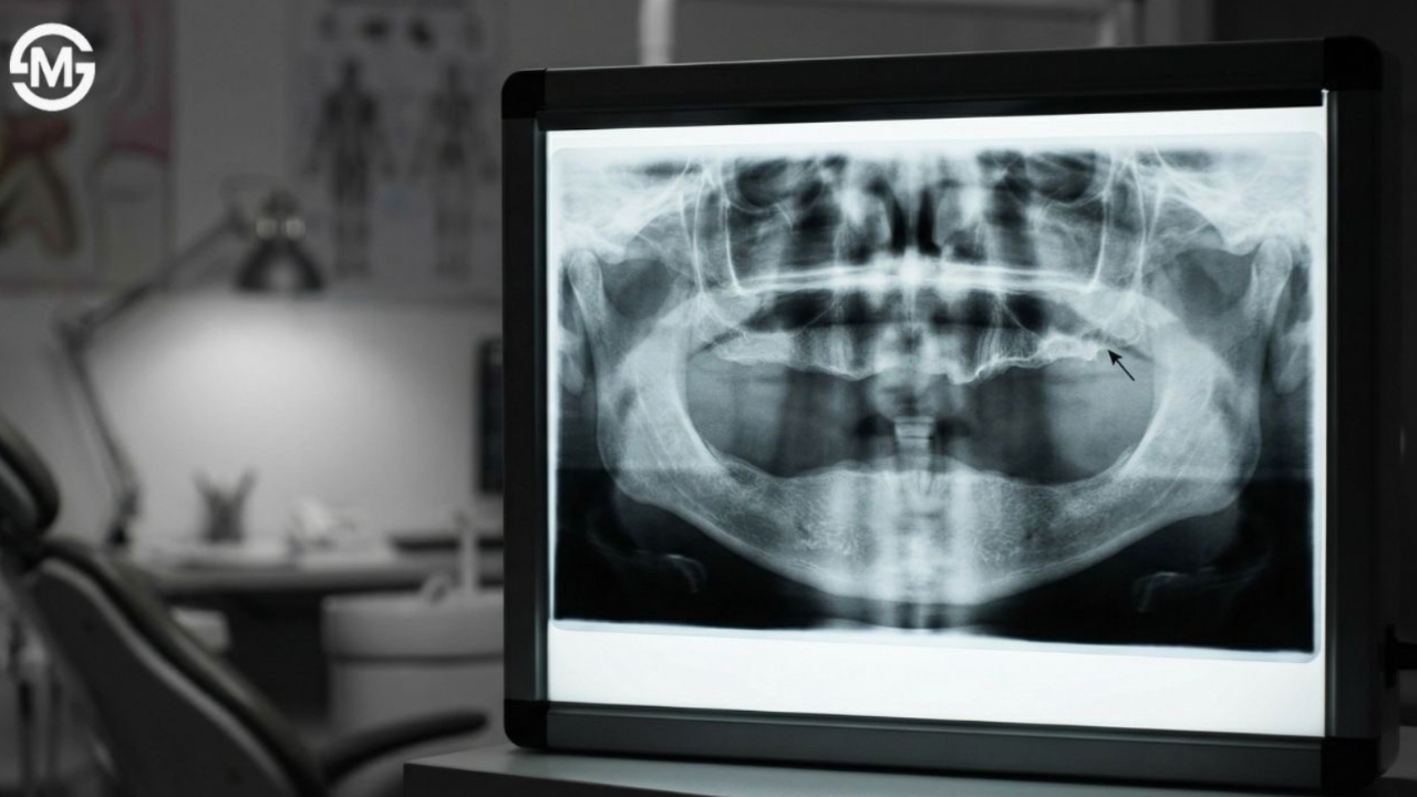
In the Mandible, the zone of keratinized tissue is typically much narrower, which demands a more conservative approach. In straightforward mandibular cases, a mid-crestal incision remains the most prudent choice, ensuring an even distribution of the limited tissue between the buccal and lingual aspects. However, in Amanda's case — after a guided bone regeneration procedure using a bone allograft, a pericardial membrane, and the patient's own platelet-rich fibrin, followed by an eight-month healing period — I utilized an apically repositioned flap during the second-stage surgery. This technique allowed me to surgically move the entire band of tissue apically, creating a wide, stable, and well-attached zone of keratinized gingiva around the emerging implant abutments.
The 15-Year Result That Changed How I Think About Surgery
Amanda, who had been told she would never have dental implants, has now maintained a stable, functional, and aesthetically excellent result for over 15 years. The bone has remained intact. The tissue has remained healthy. The implants have remained stable. When I look at her follow-up records today, the result is as good as — in some ways better than — cases I have treated in patients with ideal anatomy.
The lesson I take from Amanda's case — and from the hundreds of cases I have treated using this approach since — is that the most powerful tools in surgery are often not the newest ones. They are the ones that require us to think more carefully about what we already have. The incision is not a formality. It is a decision. And like every decision in surgery, it carries consequences that will unfold over years and decades.
I encourage every surgeon reading this to look at your next case and ask: where is the keratinized tissue right now, and where do I need it to be? The answer to that question — and the incision placement that follows from it — may be the most important surgical decision you make that day.


